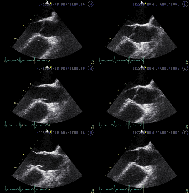
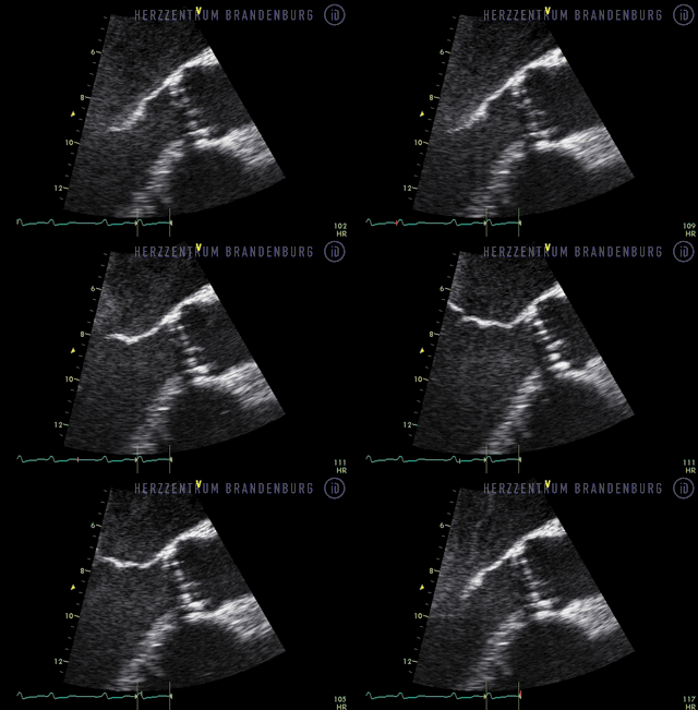
Aortic stenosis
The heart valve that separates the left ventricle from the aorta may lose its flexibility as a result of ageing or inflammation. The heart valve is no longer able to open fully, thus restricting the flow of blood.
Contact
What is aortic stenosis?
Aortic stenosis is usually associated with increasing age. The aortic valve - which separates the left ventricle from the aorta - loses its flexibility as a result of age-related or inflammatory changes. Consequently, aortic stenosis refers to a narrowing of the valve between the left ventricle and the aorta. As the valve can no longer open fully, it impedes blood flow to the rest of the body, which puts increased strain on the heart.
If left untreated for a number of years, this will result in reduced exercise capacity and heart failure. Depending on the severity of the condition, aortic stenosis may remain undiagnosed for a number of years. As a rule, treatment becomes necessary once symptoms develop, as the prognosis of symptomatic patients, and survival rates, are poor.
What are the main symptoms?
- Heart failure that may be associated with reduced exercise capacity, as well as shortness of breath during exercise or even at rest.
- Angina pectoris (e.g. as chest pain upon exertion) due to aortic stenosis causing an inadequate supply of blood to the heart muscle.
- Syncope (sudden loss of consciousness) due to arrhythmia, which develops as a result of the condition.
ECG results that show a considerable increase in the average pressure gradient across the aortic valve, are an important indicator of severe aortic stenosis (Fig. 1).
![[Translate to Englisch:] EKG-Messung - Immanuel Herzzentrum Brandenburg in Bernau - Aortenstenose [Translate to Englisch:] EKG-Messung - Immanuel Herzzentrum Brandenburg in Bernau - Aortenstenose](/assets/IKBHB/Zentren/Hezzentrum/03_Leistungsspektrum/03_01_Krankheitsbilder/immanuel-herzzentrum-brandenburg-in-bernau-aortenstenose-messung.jpg)
Diagnosis of aortic stenosis
- Auscultation (listening to the sounds of the heart using a stethoscope): aortic stenosis produces characteristic sounds in the heart, which can be detected with the help of a stethoscope.
- Echocardiography (ultrasound examination of the heart): this is the most important examination in the diagnosis of aortic stenosis, as it allows the visualization of relevant structures. The physician can use it to measure the pressure gradient across the diseased valve, an important indicator of the degree of stenosis (narrowing) present.
- Left heart catheterization (a sound probe inserted into the heart via blood vessels in the arms or legs): this procedure allows the physician to take direct measurements of the pressure gradient across the diseased valve. This is important in determining the severity of stenosis (narrowing) present, and allows the visualization of other areas that may be affected by reduced blood flow.
- Cardiac CT: at this point in time, this procedure is only of minor significance in the diagnosis of aortic stenosis.
- Cardiac MRI: at this point in time, this procedure is only of minor significance in the diagnosis of aortic stenosis.


Treatment for aortic stenosis
- Medication
- Standard treatment
- Transcatheter aortic valve implantation (TAVI)
- Balloon valvuloplasty
Medication
Medications can be used to ease the early symptoms of aortic stenosis. However, they do not have any significant impact on the future course of the disease.
Standard treatment
The standard treatment for aortic stenosis consists of the replacement of the diseased valve with a prosthetic valve (tissue or mechanical).
Transcatheter aortic valve implantation (TAVI)
Transcatheter aortic valve implantation (TAVI): this procedure is particularly suited for patients who, due to advanced age or concomitant disease, are at increased risk of complications during surgery. During this procedure, a valve, which is mounted onto a delivery catheter, is implanted via a blood vessel in the groin (transfemoral) or via an incision directly into the apex (tip) of the heart (transapical).
The first-in-man procedure was performed in 2002. Since 2007, this procedure has become increasingly widespread. In terms of experience with TAVI procedures, Germany's position is unparalleled in the world. Brandenburg Cardiac Centre is one of Germany's "High Volume Centers" and as such has significant experience with this procedure. Physicians from the Center's Cardiology and Cardiac Surgery Departments, who perform this procedure in our Center, also provide training for other physicians around the world.
Balloon valvuloplasty
While valvuloplasty (catheter-based procedure to widen the aortic valve using a balloon-catheter) can help ease symptoms in the short term, its long-term effects are limited.
Specialist publications
Rotational angiography for pre-interventional imaging in transcatheter aortic valve implantation
Meyhoefer J. MD, Ahrens J. MD, Neuss M. MD, Hölschermann F. MD, Schau T. MD, Butter C, MD., Cath Cardiovasc Interv. 79:756–765 (2012)
Leitlinien zu Aortenklappen [Guidelines on aortic valves]
Deutschen Gesellschaft für Kardiologie – Herz- und Kreislaufforschung e.V.
leitlinien.dgk.org
ESC Working Group on Valvular Heart Disease Position Paper
European Heart Journal
eurheartj.oxfordjournals.org
Transcatheter Aortic-Valve Implantation for Aortic Stenosis in Patients Who Cannot Undergo Surgery
The New England Journal of Medicine
www.nejm.org
Where can I find out more about aortic stenosis?
General information
Deutsche Herzstiftung e.V. (German Heart Foundation)
www.herzstiftung.de
