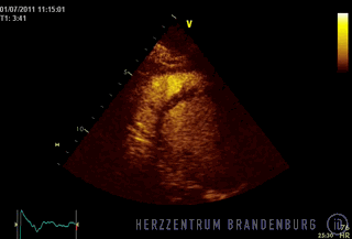
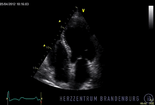
Echocardiography
An echocardiogram is a very quick and non-invasive imaging technique that allows a thorough examination of the patient's heart without causing any trauma or discomfort to the patient. This technology allows a detailed assessment of the size of the various chambers of the heart, as well as providing a clear view as to how well the heart and the various heart valves are working. It can also be used for the quick and accurate detection of heart defects.
Contact
Echocardiography
Patients of the Brandenburg Heart Center have access to the full range of echocardiology equipment and procedures available. In addition to 2D imaging and conventional Doppler techniques, we also offer modern techniques such as tissue Doppler imaging and speckle tracking echocardiography, as well as contrast echocardiography and 3D echocardiography.
Heart disease that becomes apparent in response to physical stress can be diagnosed using stress echocardiography.
Every year, we perform approximately 6000 cardiac ultrasound examinations.
Transesophageal echocardiography (TEE)
Transesophageal echocardiography is a cardiac ultrasound examination that is performed via the esophagus and is used for the accurate assessment of heart valve function and congenital heart defects. The ultrasound probe is inserted through the patient's mouth and advanced down the throat. The patient will be asked to swallow to help the probe "go down". The patient's throat will be numbed with a local anesthetic and, in most cases, the patient will also be sedated to ensure that the procedure causes as little discomfort as possible. The probe is then advanced through the esophagus and into the stomach. Due to the fact that the heart is positioned directly in front of the esophagus, and the ultrasound does not have to travel through lung tissue before reaching the heart, this technique results in much better image quality. The procedure usually takes between 10 and 15 minutes and can be performed on an outpatient basis.
As this technique allows a thorough examination of the atria and heart valves, it is also used to assist navigation during catheter-based interventions involving the heart valves. The technique is used during many types of cardiac surgery where it can monitor progress throughout the procedure. Although the procedure can be performed on an outpatient basis, this involves sedating the patient. When the patient is ready to be discharged, they will therefore need to be accompanied home by a responsible adult.
Ultrasound scanning
In our hospital, all diagnostic ultrasound scans are performed by experienced operators and using the latest in modern technology. There are two ultrasound suites available, as well as a radiology reading room with two workstations. These can be used to access and review a patient's complete ultrasound records, thus allowing physicians to monitor disease progress by comparing new data with those from previous ultrasound scans.
Team meetings
These work stations are also used as part of the regular team meetings with our colleagues from Cardiac Surgery and Interventional Cardiology, and allow us to make team-based decisions regarding treatment strategies and treatment planning, including for instance decisions on the type of surgical or catheter-based procedure that might be best suited to treat a particular patient's heart valve disease.
3D echocardiography
3D cardiac ultrasound offers new possibilities in the field of echocardiography. In a process that takes only a matter of seconds, ultrasound technology is used to produce a 3D visualization of the entire heart.
This enables physicians to make use of echocardiography-guided navigation during the highly complex, minimally-invasive catheter-based procedures they perform in our catheterization laboratory. These types of procedures include left atrial appendage closure (using the Watchman device), paravalvular leak repair after prosthetic valve implantation, percutaneous and transapical implantations of prosthetic aortic valves, the MitraClip procedure and the treatment of congenital heart defects using septal umbrellas.

How does a 3D echo work?
- A series of 2D ultrasound images are taken simultaneously and then used to reconstruct a 3D model which can also be rotated.
- Images can be obtained from different imaging planes at the same time. In the past, this would only have been possible if done in sequence, and would have resulted in a time-consuming process involving repeated repositioning of the transducer (triplanar, biplanar imaging).
- The 3D data set can be used to produce volume-rendered images, which allow the physician to obtain fairly accurate measurements of the size of the ventricles, and make it possible to closely monitor disease progress and detect even minor changes.
- Just as is the case with cardiac CTs and cardiac MRIs, cardiac echocardiology using multislice technology can also be used to obtain cross-sectional images.
- The operator reviews and analyses the data and makes a diagnosis. Even after the procedure, the operator can retrieve the dataset to produce further images as and when required.
- The ability to reconstruct the heart in this manner makes it much easier for interventional cardiologists and cardiac surgeons to involve a patient in an interactive discussion about the details of their case, and also helps to facilitate the interdisciplinary decision-making process.
What is speckle tracking?
Speckle tracking involves the monitoring of tissue changes using the automatic tracking of characteristic patterns inside the heart muscle (which might be likened to star constellations). The changes thus observed can then be used to determine the degree of tissue deformation (strain) inside the heart muscle, and the speed at which such deformations occur (strain rate). This allows even subtle changes to be detected and provides information on the functional status of even very small areas of the heart muscle. This technique is used as part of very specific investigations in patients with congenital heart disease or an inflammation of the heart muscle (myocarditis).
It is also an important tool in assessing the potential benefit a patient might derive from biventricular stimulation (cardiac resynchronization therapy).
Es hat auch hohe Bedeutung zur Einschätzung, inwieweit ein Patient von einer biventrikulären Herzstimulation (cardiale Resynchronisationstherapie) profitiert.
What is stress echocardiography?
Aortic valve stenosis - a narrowing of the aortic valve due to e.g. calcification - is the most common type of valve disease affecting elderly people. In many cases, it is difficult to determine the best point at which to perform surgery to treat the diseased valve and, particularly in patients with aortic valve stenosis with low output (low-gradient aortic stenosis), stress echocardiography can help us to make the right decision.
- Stress echocardiography is used to diagnose heart disease that becomes apparent in response to physical stress. It is primarily used patients with suspected myocardial ischemia, and to identify areas of viable myocardial tissue in patients with known coronary heart disease.
- Stress is usually drug-induced but in some cases, where more targeted investigations are required, the patient may instead be asked to exercise on an exercise bike.
- When drugs are administered, these make the heart beat faster and harder, simulating physical stress.
- Global strains and regional deformations of the heart are assessed by acquiring images from different imaging planes (e.g. apical four chamber view, parasternal short axis view).
- Disturbances in wall motion observed under stress are indicative of impaired blood flow (ischemia).
- In patients where the images obtained are of a very low quality, ultrasound contrast agents can be used to improve left ventricular opacification. This often leads to a clearer view of the left ventricle, and in turn facilitates the assessment of both global and regional heart function.