The anatomy of the heart
The heart is one of the most important, complex and fascinating organs in the body. This section will tell you more about the heart and the work it does for you every day of your life.
Contact
What is the heart and how does it work?
The heart is a hollow, muscular organ whose rhythmic contractions lead to the movement of blood and lymph around the body, thus ensuring organ perfusion. Ibn al-Nafis (1213-1288), an Arab physician, was the first person to provide an anatomically-correct description of the heart, while the English physician William Harvey (1587-1657) is credited with being the first person to demonstrate that the heart's pumping action results in the movement of blood around the body.
The heart is roughly the size of a fist, with its shape resembling that of a cone with rounded edges, and with its tip (apex) pointing downwards and slightly towards the front and left. In the human body, the heart is located behind the sternum (breastbone) and slightly to the left of the midline. At approximately 0.5% of a person's body weight, a healthy heart averages between 300 and 350g.

Layers of the heart wall
The heart is completely enclosed by the pericardium: a sac made of connective tissue. Beneath the pericardium is the epicardium, a layer of the heart that contains adipose tissue (epicardial fat) and which encloses the coronary arteries. Beneath the epicardium is the myocardium - the actual heart muscle. On the inside, the heart is lined with a thin membrane, the endocardium.
Chambers of the heart and blood vessels
On the inside, the heart is lined with a thin membrane, the endocardium.
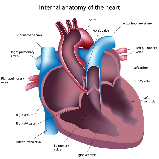
The human heart has four chambers, two upper chambers (atria) and two lower chambers (ventricles). The atria and ventricles are separated by the septum, while the heart valves act as one-way valves - ensuring that blood flow through the heart is only in one direction. Blood is transported away from the heart and towards the organs via a single artery. Blood returning from the organs back to the heart does so via a single vein.
The superior and inferior vena cava, which feed into the right atrium (RA), return oxygen-depleted blood from the systemic circulation to the heart. When the ventricles contract, the tricuspid valve (the valve between the right atrium and the right ventricle) prevents the blood from flowing back into the atrium. Blood is then pumped out of right ventricle (RV) and towards the lungs, with the pulmonary valve preventing any back-flow of blood into the right ventricle. The pulmonary artery feeds oxygen-depleted blood into the pulmonary circulation.
Once the blood has been enriched with oxygen, the pulmonary circulation returns it into the left atrium (LA) via pulmonary veins, of which there are normally four in the human body. From here, the blood enters the left ventricle (LV) via another cusped valve - the mitral valve. Blood flow out of the left ventricle is via a further cusped valve - the aortic valve.
Coronary arteries
The coronary arteries, which supply the heart with oxygen, exit the aorta just above the level of the aortic valve. There are two coronary arteries in the body, the right coronary artery (RCA) and the left coronary artery (LCA). The main stem of the left coronary artery (LMCA) divides into two main branches, the left circumflex artery - ramus circumflexis (RCX) - which supplies the side of the heart, and the left anterior descending artery - or ramus interventricularis anterior (RIVA) - which supplies the anterior wall of the heart.
