Pulmonary vein isolation for atrial fibrillation
Atrial fibrillation can be caused by electrical signals that originate in the area around the pulmonary veins. If this is the case, catheter ablation can be used to isolate the pulmonary vein from the left atrium. This is referred to as pulmonary vein isolation.
Contact
What is pulmonary vein isolation?
Pulmonary vein isolation is a minimally-invasive catheter-based electrosurgical technique for the treatment of atrial fibrillation. The purpose of treatment is to prevent a recurrence of the symptoms of atrial fibrillation. Pulmonary vein isolation should be considered in patients who remain symptomatic despite optimal pharmacological therapy.
Pulmonary vein isolation prevents electrical signals from the pulmonary veins from reaching the left atrium. The pulmonary veins are responsible for producing frequent electrical signals (triggers) that cause atrial fibrillation. Ablation can isolate the triggers produced in the pulmonary veins, thus stopping them from reaching the atrium and triggering atrial fibrillation.

What happens during pulmonary vein isolation?
Catheter ablation offers a potential treatment option in patients with symptomatic paroxysmal atrial fibrillation.

Prior to the procedure
Certain tests may be required prior to the actual procedure. These will provide the technical data necessary for the procedure, as well as helping to make the procedure safe. The decision as to which types of tests are required is made on a case-by-case basis.
- Trans-thoracic (TTE) and trans-esophageal (TEE) echocardiography - an ultrasound examination to establish the precise anatomy of the heart.
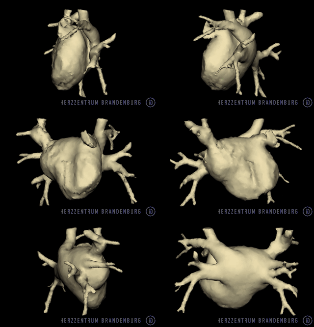
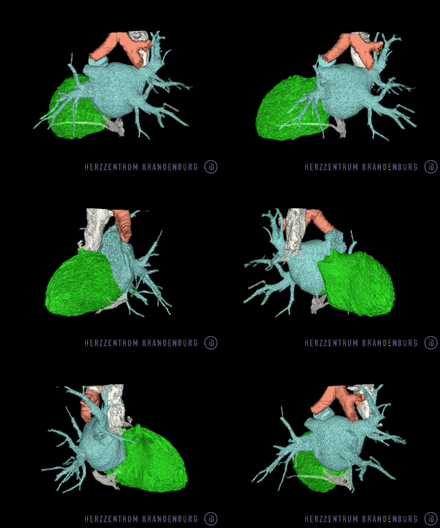
- MRI (magnetic resonance) imaging or CT (computed tomography) scanning to establish the precise anatomy of the left atrium. Amongst other things, these imaging data will be used to produce a 3D reconstruction during the procedure.
- Significant disruptions to normal blood flow through the heart need to be ruled out. If in doubt, left heart cardiac catheterization (coronary angiography) may be required
The examination takes place in a specially equipped catheterization laboratory - our electrophysiology laboratory. Prior to undergoing the actual procedure, you will be connected to a number of different monitors (e.g. ECG). This allows continuous monitoring of your heart function throughout the procedure.
During the procedure
The procedure is performed using local anesthesia. In most cases, additional drugs will be administered that also induce sleep (procedural sedation).
A catheter is inserted into the femoral vein and advanced toward the heart. It enters the right atrium via the vena cava, passing through the septum into the left atrium.
Once the left atrium and the pulmonary veins have been visualized using contrast agents, a special computer-guided system is used to produce a 3D mapping image of the left atrium. This image is then used as a guide throughout the remainder of the procedure.
Once the relevant area has been successfully ablated, the procedure is often interrupted for a considerable period of time in order to determine whether the ablated tissue is likely to recover. At the end of the procedure, the catheters are removed.
The procedure normally takes between 2-4 hours.
Robotic catheterization procedure (Sensei X - Hansen Medical)
In our hospital, we have the option of performing this procedure using a robotic navigation system. This allows the physician to operate the catheters remotely, from a control room, reducing radiation exposure for both the patient and the operator. In addition, catheter stability is improved, which may make difficult-to-reach areas more accessible. The decision as to whether robotic navigation is a viable option must be made on a case-by-case basis.
After the procedure
The patient remains in the catheterization laboratory while a pressure bandage is being applied, and will need to remain in bed for a number of hours after the end of the procedure. It will be necessary for the patient to be closely monitored for at least one day after the procedure.
Discharge from hospital
It is usually possible for patients to be discharged from the clinic a day or two after the procedure.
At home
After discharge, you should try to avoid physical activity for a duration of one week.


Where can I find out more about pulmonary vein isolation?
General information
Deutsche Herzstiftung (German Heart Foundation)
Deutsche Gesellschaft für Kardiologie (German Cardiac Society)
dgk.org
Kompetenznetz Vorhofflimmern (The German Competence Network on Atrial Fibrillation, AFNET)
kompetenznetz-vorhofflimmern.de
Sources
Leitlinien, Deutsche Gesellschaft für Kardiologie - Katheterablation [Guidelines, German Cardiac Society - Catheter Ablation]
