Pacemakers
Pacemakers are implantable devices that are used to monitor and treat patients with bradycardia (slow heart rhythm). The implantation of pacemakers is a routine surgical procedure.
Contact
What is a pacemaker?
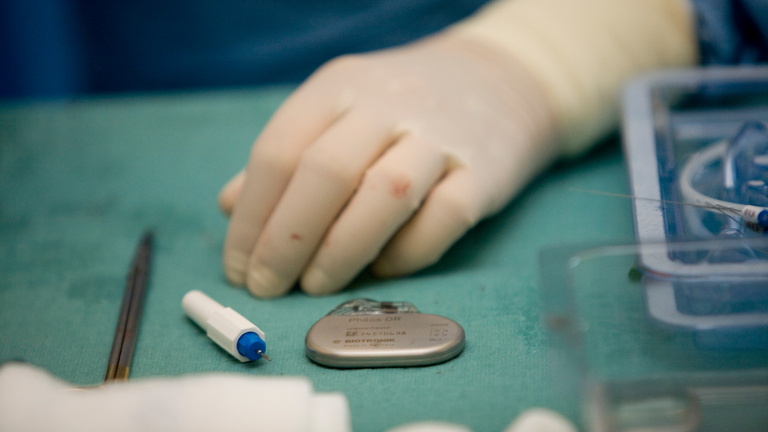
A pacemaker is an approximately matchbox-sized device that is used to monitor and treat bradyarrhythmias (slow heart rhythms).The device uses electrical impulses to stimulate the heart muscle in a regular and rhythmic manner, resulting in rhythmic contractions of the heart.
Development of pacemakers
On 8. October 1958, Arne Larsson became the first patient to be fitted with a fully-implantable pacemaker. The procedure was carried out by Rune Elmqvist, an engineer working at Siemens-Elema, and Åke Senning, a Swedish cardiologist.
Since then, pacemaker implantation has become a routine procedure. In 2009, a total of approximately 70,000 pacemaker implantation procedures were performed in Germany.
What conditions can be treated using a pacemaker?
- Bradycardia (slow heart rhythm) due to:
Sinus node disease
Disorders affecting the AV node (referred to as 2nd and 3rd degree AV block)
Disorders of the heart's conduction system that affect both impulse generation and impulse propagation - Slow atrial fibrillation
Treatment step-by-step
Pacemaker implantation is a routine surgical procedure, with more than 220 pacemakers being implanted at our Heart Center every year.
If a patient is diagnosed with a type of bradyarrythmia (slow heart rhythm), the implantation of a pacemaker may become necessary. The decision as to whether the patient receives a single chamber, dual chamber or biventricular pacemaker (i.e. with one, two or three leads) will depend on the nature of the patient's underlying disease and heart rhythm, and will be made in accordance with the recommendations and guidelines set by the German Cardiac Society (Deutsche Gesellschaft für Kardiologie).
Patients with slow atrial fibrillation are fitted with a single chamber pacemaker, i.e. a pacemaker with one lead to stimulate the ventricle. Patients with intermittent sinus rhythm are fitted with a dual chamber pacemaker, i.e. a device with two leads, which can help to maintain the synchronized action of the atria and ventricles. Patients with decreased ventricular function may need to be fitted with a biventricular pacemaker, i.e. a device with three leads that ensures optimal synchronization of the right and left ventricles.
The implantation of the control unit is performed under local anesthesia, and involves a small incision approximately 5cm in length being made below the collar bone on either the left or right side. The leads (with electrodes at their tips), which connect the pacemaker to the heart, enable it to measure and analyze the heart's electrical activity and, if required, deliver localized electrical impulses to stimulate the heart muscle, thus ensuring that the heart continues to beat.
Follow-up treatment
Regular follow-up appointments either via the outpatient department or a local specialist; home (remote) monitoring where appropriate.
